|
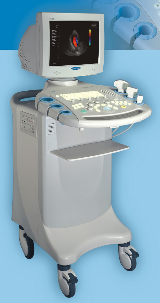
Digital Color Doppler (ADW2000)
PRODUCT'S INFORMATION:
Model ADW2000 is a mobile ultrasound scanner whose
magnificent performances define its technology superiority
over other available ultrasound machines in the market. This
new fully digital ultrasonography machine is ideal for high
volume institution that requires a state of the art
ultrasonography technology in the market without breaking the
bank. The high-precision digital imaging technology that the
unit is armed with works harmoniously with the system's
state of the art software packages to give you the best
image that you can ever expect from any ultrasound machine
including, but not limited to, live-picture like
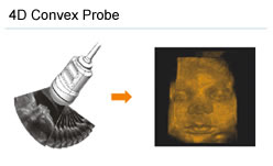
4D
images that give the sonographer and his or her patient a
clear picture of the unborn baby as if it just had its
pictures taken postnatal.
This unit will definitely give your facility the long
awaited satisfaction in the sonography imaging studies. It
provides you crisply sharp images of structures with excellent performances in varieties of
ultrasonography imaging studies in various fields including,
but not limited to, Obstetrics and Gynecology, Cardiology
and Urology.
The important package
that comes with our products, which many distributors or
manufacturers across the globe cannot guarantee, is our
eighteen month-post sales' manufacturer warrantee that
ensures that our customers are fully protected against any
financial responsibility in the non-user's required maintenances that
are required for hitch free performances of our products
during the first eighteen months after purchasing any of our
products. If any product you buy from us malfunctions within
the first eighteen months of its life in serving you, just
contact us and we will service or replace it promptly
without any financial responsibility to you. In addition to
this, we have many other warrantee packages that cover our
products.
Special Features:
1) High-precision digital imaging technology.
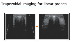
2) THI (Tissue Harmonic Imaging), real-time compound
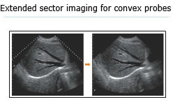
imaging, trapezoidal imaging, extended sector imaging.
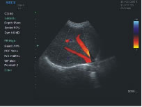
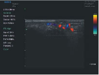
3) Color Doppler Flow Imaging (CDFI), spectrum Doppler.
4) Extensive software packages for various measurement and
clinical diagnostic calculation function.
5) Extensive series of super broadband, high density probes,
like phased array, 4D.
6) Built-in workstation.
7) Powerful image and file management systemic PACS systems
and DICOM3.0 connection.
State-of-the-art Imaging technology presents high quality
images:
High-precision digital imaging technology displays fine
tissue structure.
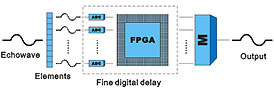
Digital beam forming technology:
Accurate beam forming, signal processing, digital image
procurement and processing ensure images with clear-cut edge
and no distortion.
The following is a fine digital display of the unit:
You can choose to extend
an image from a trapezoidal format to a wider view:
The unique
Beam Sector Extension 1 function allows
observing viscus in a wider scope without dual
concatenation:
|
 |
 |
|
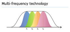
All probes are super-broadband with over 70%
relative bandwidth. Different frequency ranges are
available for each probe on different clinical
requirements. |
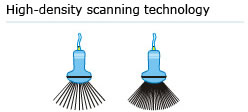
Real-time Dynamic high-density beam scanning |
| |
|
 |
 |
|
Realtime 3D/4D imaging to acquire true three
dimensional information automatically and quickly.
|
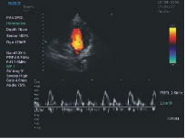
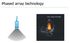
Equipped with phased array probes - the system is
suitable for cardiac diagnosis through rib gap to
acquire a wide-angle image. |
Extensive Series of wide band probes and software
packages:
The Apogee 3500 is available with many high-density,
super broadband and multi-frequency probes, such as
convex, micro-convex, linear, vaginal, rectal and phased
array probes, which are widely applied for different
clinical diagnoses, including abdomen (liver, kidney,
gall-bladder, pancreas), gynecology (uterus, ovary),
obstetrics (early pregnancy, basic OB, complete OB,
multi gestation, fetal echo), cardiology (adult and
pediatric cardiology), small parts (thyroid,
galactophore, testicles, neonate), peripheral vascular
and prostate.
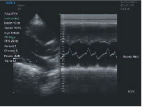
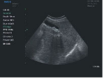
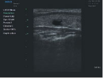
The followings are
some of the images taken with ADW2000 sonogram from their
respective body structures:
|